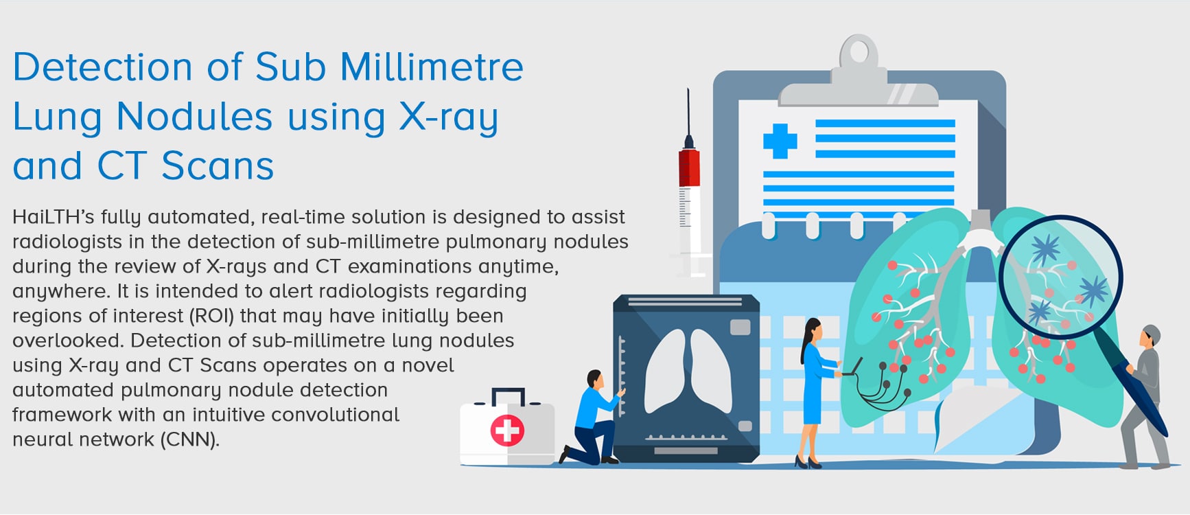
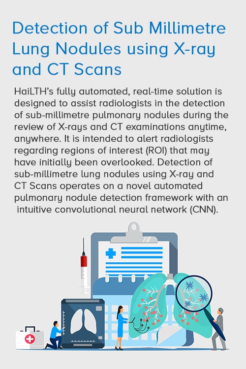
Progress and Prospects of Using AI for Chest Radiographs
On one hand, the rapid rise in the use of chest imaging has led to increased incidental lung nodule findings on imaging studies. These minute nodules have long been proved problematic for follow-up. On the other, the ever-growing workload of radiologists often results in misses, delaying diagnoses of patients. In such a scenario, using a solution based on deep learning technology and specifically trained using millions of images helps in the following:
sub-millimetre lung
nodules quickly and easily
high-quality reports
anytime anywhere to
increase physician
satisfaction and patient care
incidental pulmonary nodules
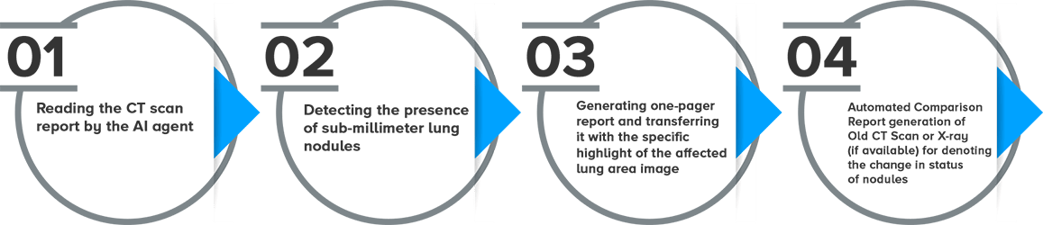
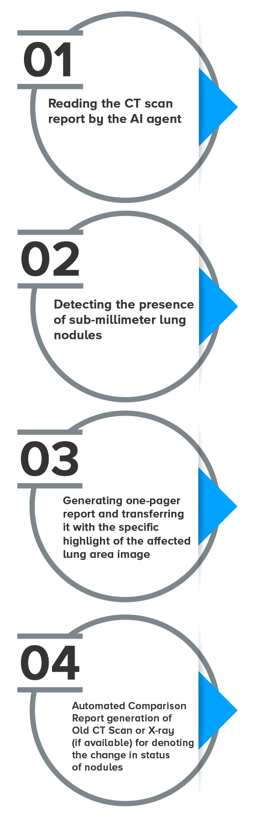
Four Steps to Detection and Management of Sub Millimetre Lung Nodules
Driving Care at Scale with AI

According to the World Health Organization (WHO), lung cancer is the most common cancer with approximately 2.09 million new diagnoses worldwide each year. The disease is also responsible for most cancer deaths at around 1.76 million. More than 80% of lung cancer patients are likely to survive for at least a year when diagnosed at the earliest stage compared to about 15% of those diagnosed at an advanced stage of lung cancer. Thus, the earlier lung cancer is detected the better chance a patient has of surviving the disease.
HaiLTH leverages RAD 365 AI-based platform to enhance radiologists’ ability to detect pulmonary nodules while reducing chest X-ray, CT scan interpretation times and increasing the efficiency, quality, and precision in reporting lung abnormalities.
According to the World Health Organization (WHO), one in six deaths is due to cancer. With about 160,000 deaths in 2018, lung cancer remains one of the most common cancers. Lung cancer screening using low-dose computed tomography has been shown to reduce mortality by 20–43%.
